

From Redness to Craters: Understanding the Four Stages of Bed Sores
- Brett Leitner
- May 16, 2023
- 4 min read

A pressure ulcer, also known as a bed sore, is a localized injury to the skin and underlying tissue that usually develops over a bony protrusion or a pressure point. It occurs when unrelieved pressure and excessive friction or shearing forces damage the soft tissue, impede blood flow, and cause the cells to die or break down. The resulting wound can vary in severity and depth, from a mild redness or blister to an open sore or a deep cavity that exposes muscles, bones, or organs.


Pressure ulcers are common among people who are bedridden, wheelchair-bound, or have limited mobility, such as those who are elderly, terminally ill, or have neurological disorders or spinal injuries. According to the National Pressure Ulcer Advisory Panel (NPUAP), pressure ulcers affect approximately two to three million people in the United States each year, and the prevalence can vary from 10 to 33 percent in hospitals, nursing homes, and long-term care facilities.
While pressure ulcers can develop on any part of the body, there are certain areas where they are more common.
Buttocks: The buttocks are the most common site for pressure ulcers. This may be due to their bony prominence and the fact that the weight of the body is primarily distributed across this area. This constant pressure can lead to damage to the skin and underlying tissues.
Hips: Hips are another common site for pressure ulcers in individuals who spend a lot of time in bed or sitting in a chair. Pressure sores can develop along the hip bone area when there is prolonged pressure.
Heels: Pressure sores on the heels are common in people who are immobilized and are unable to move their feet. Pressure ulcers may develop on the heels due to pressure and friction caused by shoes and boots.
Shoulder blades: If a person is lying on their back or sitting in a wheelchair for extended periods, they may develop pressure sores on their shoulder blades. This is because the bones protrude from the skin, making them a target for pressure and friction.
Elbows: Elbows are another commonly affected site for pressure ulcers, particularly in people who spend a lot of time leaning on their elbows or are unable to move their arms frequently.
Back of the head: Pressure ulcers may also develop on the back of the head, particularly in individuals who spend most of their time lying in bed. This can be due to the pressure of the head against a mattress or pillow.
It is important to note that while these areas are the most commonly affected, pressure sores can occur on any part of the body. Healthcare professionals must be vigilant in assessing for pressure sores and should always assess and evaluate the patient's risk factors for developing pressure ulcers. Proper prevention and management of pressure ulcers can help to avoid the development and worsening of these wounds and promote patient wellness and safety.
The causes of pressure ulcers can be attributed to several factors, including immobility, incontinence, poor nutrition, dehydration, medication side effects, and impaired sensory perception. The risk of developing pressure ulcers can be assessed based on the patient's age, health status, activity level, and exposure to pressure and friction.
The National Pressure Ulcer Advisory Panel (NPUAP) has established a classification system that differentiates four stages of pressure ulcers, ranging from mild to severe. Each stage represents a unique level of tissue damage and requires a different approach to treatment:
Stage 1: In the initial stage of a pressure ulcer, the skin is not broken, but there may be some discoloration or a persistent area of redness that does not go away when pressure is relieved. This may indicate that damage has occurred to the underlying tissue. The affected area may feel warmer to the touch than surrounding skin and is often painful or tender.
Stage 2: In stage 2, the skin has become broken and a shallow open wound has developed. The wound may look like a blister or an abrasion, and may be surrounded by more extensive damage to the skin. The wound may still be painful or tender at this stage, and the affected area may feel warm or hard to the touch.
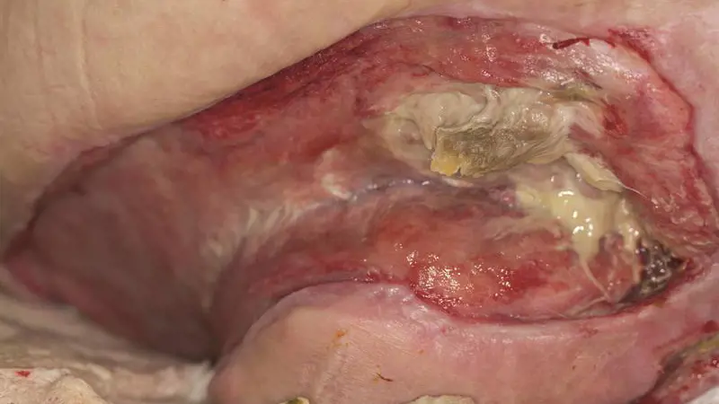
Stage 3: At this stage of a pressure ulcer, the wound has deepened and expanded. The damage has now extended beyond the skin to affect the subcutaneous fat layer. The wound may appear as a deep crater and may involve significant tissue loss. It may or may not be painful, as nerve damage may have occurred.
Stage 4: In this final stage of a pressure ulcer, the wound has extended deep into the underlying tissue and may affect muscles, tendons, and even bones. The wound may appear as a large crater and may involve significant tissue loss. The wound is often painful, as nerve damage has occurred.
The prevention and treatment of pressure ulcers involve a comprehensive approach that includes identifying and managing the underlying risk factors, providing supportive surfaces and positioning devices, maintaining skin hygiene and moisture, and using appropriate wound care products and modalities. The NPUAP has also established guidelines and best practices for the prevention and treatment of pressure ulcers, which involve regular skin assessments, documentation of ulcer status, communication among healthcare providers, and patient education and participation.
Pressure ulcers are a serious and preventable complication of prolonged immobility and other risk factors that can cause significant pain, disability, and morbidity. It is essential to recognize the signs and symptoms of pressure ulcers, assess the risk factors, and implement timely and appropriate interventions to prevent and treat this condition. Healthcare professionals have a responsibility to be knowledgeable about pressure ulcer prevention and management and to advocate for our patients' wellness and safety.
Understanding the different stages of pressure ulcers is essential in providing appropriate and timely treatment for patients. It is important to recognize the signs and symptoms of each of the four stages of pressure ulcers, assess the risk factors, and implement timely and appropriate interventions to prevent and treat this condition.
Sources:
National Pressure Ulcer Advisory Panel. Pressure ulcers: incidence, economics, and risk assessment. https://www.npuap.org/wp-content/uploads/2018/02/Pressure-Ulcers-Incidence-Economics-Risk-Assessment-2018-02-08-2.pdf
National Institute on Aging. Bed sores (pressure ulcers). https://www.nia.nih.gov/health/bed-sores-pressure-ulcers
Mayo Clinic. Pressure injuries. https://www.mayoclinic.org/diseases-conditions/pressure-injuries/symptoms-causes/syc-20355897
Wound, Ostomy and Continence Nurses Society. Guideline for prevention and management of pressure injuries. https://cdn.ymaws.com/www.wocn.org/resource/resmgr/publications/wocs_guideline_pressure_ul.pdf
WebMD. Understanding pressure sores symptoms. https://www.webmd.com/skin-problems-and-treatments/understanding-pressure-sores-symptoms


